

Scientist of the Day - Max Perutz

Max Perutz with his 1959 low-resolution, balsa-wood model of hemoglobin, MRC Laboratory of Molecular Biology, Cambridge (mrc-lmb.cam.ac.uk)
Max Perutz, an Austrian/British molecular biologist, was born May 19, 1914, in Vienna. At the Cavendish Laboratory at Cambridge, in the late 1930s, he began pursuing the molecular structure of hemoglobin (or haemoglobin, as the British call it). The only tool at hand for probing the structure of a molecule was X-ray crystallography, which had been invented by Max von Laue in 1912 and used immediately by such experimental physicists as William Bragg and his son Lawrence. Trying to come up with a three-dimensional picture of hemoglobin was an absurd challenge; the Braggs had worked on such problems as the crystalline structure of sodium chloride, which has 2 atoms and a molecular weight of 58. Hemoglobin, it would turn out, contains 10,000 atoms, has a molecular weight of around 65,000, and consists of four separate chains of proteins bound to what are called heme groups, where the iron atoms are found and where the oxygen gets bound. How could one possibly work out the arrangement of all those atoms?

Nobel Prize winners for 1962; Max Perutz is second from left; other winners are, left to right, Maurice Wilkins, Francis Crick, John Steinbeck, James D. Watson, John Kendrew (achievement.org)
But hemoglobin can be crystallized (a necessity when using X-ray crystallography), and it is thoughtfully colored red, which means the crystals are visible and can be sorted by hand under the microscope. Perutz worked on the problem for decades, interrupted by World War II, and made his big breakthrough in 1953, when he discovered that adding heavy atoms like mercury to the hemoglobin would change the structure and alter the X-ray interference patterns in meaningful ways, allowing the three-dimensional structure to be gradually inferred. Finally, in 1959, Perutz unveiled what he called a low-resolution model of hemoglobin, which in fact was carved out of balsa wood. The photograph (first image) is a little odd, because it shows an 85-year-old Perutz posing with a model he made fully 40 years before, but I like it because it shows the model in color, unlike most earlier photographs, so one can see the four protein chains (two in white, two in black), and two of the four heme groups, in red. The model, I am told, still polices the entrance to the MRC Laboratory of Molecular Biology at Cambridge.
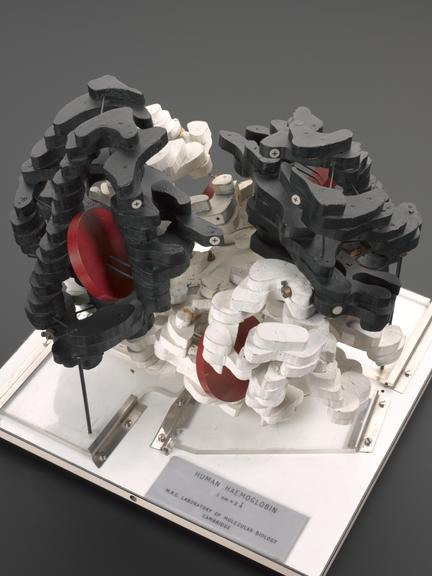
Low-resolution hemoglobin model of 1967, built by Max Perutz, Science Museum, London (sciencemuseumgroup.org.uk)
The model may have been low-res and incomplete, but it was good enough for a certain Swedish committee, which awarded Perutz the Nobel Prize in Chemistry in 1962 (which he shared with his student John Kendrew, who had worked out in a similar fashion the molecular structure of myoglobin; see our post on Kendrew). The Nobel committee cleverly arranged that the Prize in Physiology for that same year would go to James Watson and Francis Crick, for their discovery of the structure of DNA. Both achievements came out of the Cavendish Lab at Cambridge. In one of the official photographs of the 1962 Nobelists (second image), the shortish Perutz is second from left, with Kendrew at far right. Watson is next to Kendrew, and Crick is standing next to a very distinguished-looking John Steinbeck at the center, who was possibly feeling somewhat uncomfortable amidst this bevy of biologists and chemists. We have used this photograph more than once in these posts, for not only is it incredibly sharp and clear, it has as many distinguished subjects as you are likely to cram into a single close-up frame.

The high-resolution model of hemoglobin, 1968, by Max Perutz, MRC Laboratory of Molecular Biology, Cambridge (sciencephoto.com)
In 1967, Perutz built another low-resolution model, with four hinged parts so you could open it up (third image). This version is on display in the Science Museum, London.
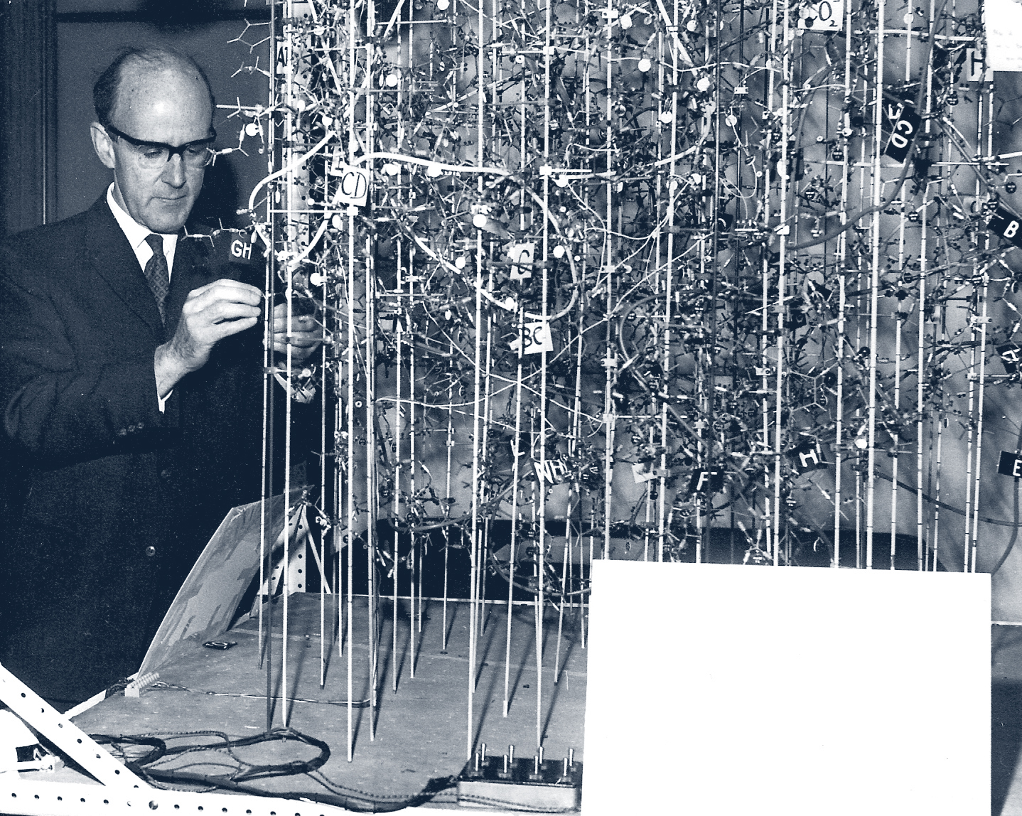
Max Perutz with his high-resolution model of hemoglobin, undated photograph, Max Perutz Labs, Vienna (maxperutzlabs.ac.at)
Perutz finally completed his high-resolution model of hemoglobin in 1968, and he was then able to show not only what hemoglobin looks like, but how it works (fourth image). The full hemoglobin model is quite something to behold. The little white rectangle in the right foreground contains a model of sodium chloride, the structure of which the Braggs had worked so hard to determine in 1914. Another photo (fifth image) shows Perutz working on, or posing with, this model. I believe the high-resolution model is still in the possession of the MRC Laboratory of Molecular Biology at Cambridge, but I do not know if it is on public display.

Ribbon diagram of hemoglobin molecule, 2007, by Richard Wheeler, using a style pioneered by Jane Shelby Richardson, 1980s (Wikimedia commons)
I thought I would link to one final image of hemoglobin, as an incentive to writing, one day, a post on Jane Shelby Richardson. Around 1980, Richardson invented a new way of depicting complicated protein molecules, using what we now call ribbon diagrams. I thought it would be fitting to conclude this notice with a recent reincarnation of a hemoglobin molecule, ribbon style (sixth image). It may not be any easier for the layperson to understand, but it has a certain elegance that a tinker-toy-style reconstruction lacks. I do not know whether Max Perutz would have been pleased with this stylish rendition of a molecular structure that he worked thirty years to determine. Given his puckish demeanor in all his photographs, he probably would have been delighted.
As with the post on Kendrew, my principal source on Perutz was Designs for Life: Molecular Biology after World War II, by Soraya de Chadarevian (Cambridge Univ. Pr., 2002). Chadarevian also wrote an insightful article on “Models and the making of molecular biology” with additional info on Perutz and Kendrew, which appeared in a book called Models: The Third Dimension of Science (Stanford Univ. Pr., 2004), which we have in the library.
William B. Ashworth, Jr., Consultant for the History of Science, Linda Hall Library and Associate Professor emeritus, Department of History, University of Missouri-Kansas City. Comments or corrections are welcome; please direct to ashworthw@umkc.edu.


